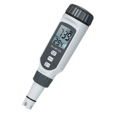
Fluorescence microscope related knowledge
The basic principle of confocal fluorescence microscopy: a point light source is used to irradiate the specimen, and a well-defined small light spot is formed on the focal plane. composed of splitters. The beam splitter sends the fluorescence directly to the detector. There is a pinhole in front of the light source and the detector, respectively called the illumination pinhole and the detection pinhole. The geometric size of the two is the same, about 100-200nm; relative to the light spot on the focal plane, the two are conjugate, that is, the light spot passes through a series of lenses, and finally can be focused on the illumination pinhole and the detection pinhole at the same time. In this way, the light from the focal plane can be converged within the scope of the detection hole, while the scattered light from above or below the focal plane is blocked outside the detection hole and cannot be imaged. The laser scans the sample point by point, and the photomultiplier tube after detecting the pinhole also obtains the confocal image of the corresponding light point point by point, which is converted into a digital signal and transmitted to the computer, and finally aggregated into a clear confocal image of the entire focal plane on the screen .
Each focal plane image is actually an optical cross-section of the specimen, and this optical cross-section always has a certain thickness, also known as an optical thin section. Since the light intensity at the focal point is much greater than that at the non-focus point, and the non-focal plane light is filtered by the pinhole, the depth of field of the confocal system is approximately zero, and scanning along the Z-axis can realize optical tomography, forming a Observe the two-dimensional optical section at the focused spot of the sample. Combining X-Y plane (focal plane) scanning with Z-axis (optical axis) scanning, the three-dimensional image of the sample can be obtained by accumulating two-dimensional images of continuous layers and processed by special computer software.
That is, the detection pinhole and the light source pinhole are always focused on the same point, so that the fluorescence excited outside the focal plane cannot enter the detection pinhole.
The simple expression of the working principle of laser confocal is that it uses laser light as the light source. On the basis of traditional fluorescence microscope imaging, it adds a laser scanning device and a conjugate focusing device, and it is a system for digital image acquisition and processing through computer control.