The main observation method of optical microscope is fluorescence observation
Fluorescence refers to the process in which a fluorescent substance emits light with a longer wavelength almost simultaneously when it is irradiated with light of a specific wavelength (Figure 1). When light of a specific wavelength (excitation wavelength) strikes a molecule, such as those in a fluorophore, the photon energy is absorbed by the molecule's electrons. Next, the electrons transition from the ground state (S0) to a higher energy level, the excited state (S1'). This process is called excitation①. The electron stays in the excited state for 10-9–10-8 seconds, during which the electron loses some energy②. During the process of electrons leaving the excited state (S1) and returning to the ground state③, the remaining energy absorbed during the excitation process is released.
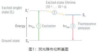
The residence time of the fluorescent molecule in the excited state is the fluorescence lifetime, which is generally on the nanosecond level, and is an inherent characteristic of the fluorescent molecule itself. Fluorescence Lifetime Imaging (FLIM), which uses fluorescence lifetime imaging technology, is called fluorescence lifetime imaging (FLIM). In addition to fluorescence intensity imaging, more in-depth functional and accurate measurements can be obtained to obtain molecular conformation, intermolecular interactions, and the microenvironment of molecules. Information that is difficult to obtain with conventional optical imaging.
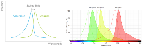
Another important property of fluorescence is the Stokes shift, the difference in wavelength between the excitation and emission peaks (Figure 2). Typically the emission wavelength is longer than the excitation wavelength. This is because electrons will lose part of their energy through the relaxation process after the fluorescent substance is excited and before releasing photons. Fluorescent substances with larger Stokes shifts are easier to observe under a fluorescence microscope.
Fluorescence Microscopy and Fluorescence Filter Cubes
Fluorescence microscope is an optical microscope that uses fluorescence properties for observation and imaging, and is widely used in various fields such as cell biology, neurobiology, botany, microbiology, pathology, and genetics. Fluorescence imaging has the advantages of high sensitivity and high specificity, and is very suitable for the observation of the distribution of specific proteins and organelles in tissues and cells, the study of colocalization and interaction, the tracking of life dynamic processes such as ion concentration changes, etc.
Most molecules in cells do not fluoresce, and to see them they must be fluorescently labeled. There are many methods of fluorescent labeling, such as direct labeling (such as using DAPI to label DNA), or immunostaining using the antigen-binding properties of antibodies, or using fluorescent proteins (such as GFP, green fluorescent protein) to label target proteins, and reversible binding. Synthetic dyes (such as Fura-2) and so on.
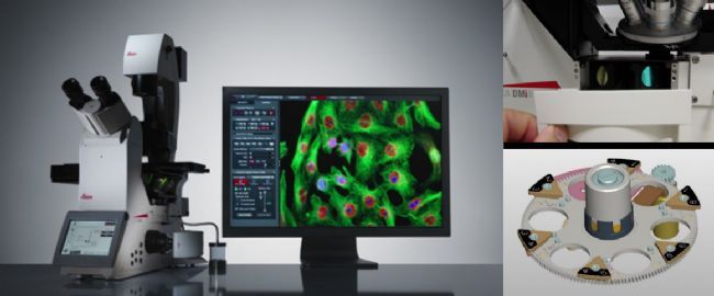
At present, the fluorescence microscope has become the standard imaging equipment of various laboratories and imaging platforms, and is a good helper for our daily experiments. Fluorescence microscopes are mainly divided into three categories: upright fluorescence microscopes (suitable for slicing), inverted fluorescence microscopes (suitable for living cells, taking into account slicing), fluorescent stereoscopes (suitable for larger specimens, such as plants, zebrafish (adult/embryo) , medaka, mouse/rat organs, etc.).
Fluorescence filter block is the core component of microscope fluorescence imaging. It consists of three parts: excitation filter, emission filter and dichroic beam splitter. It is installed in the filter wheel. For example, Leica DMi8 is equipped with a 6-position filter wheel (Fig. 3 ). The number of positions of different microscope wheels will be different, and some microscopes use filter block slides.
The filter block plays an important role in fluorescence imaging: the excitation filter selects the excitation light to excite the sample, and blocks light of other wavelengths; the light passing through the excitation filter passes through the dichroic mirror (its function is to reflect the excitation light and transmit the fluorescence), After reflection, it is focused by the objective lens, irradiates the sample, and excites the corresponding fluorescence, that is, emitted light. The emitted light is collected by the objective lens, passes through the dichroic beam splitter, and reaches the emission filter. As shown in Figure 4: the excitation wavelength is 450-490nm, the dichroic mirror reflects light shorter than 510nm, transmits light longer than 510nm, and the receiving range of emitted light is 520-560nm.