Microbial microscope direct counting method
Experimental steps (microbial microscope direct counting method)
1. Dilution
Dilute the suspension of brewing yeast appropriately. If the bacterial solution is not concentrated, it is not necessary to dilute it.
2. Microscopic examination and counting room
Before adding samples, perform a microscopic examination of the counting chamber of the counting board. If there is dirt, it needs to be cleaned before counting.
3. Add samples
Cover the clean and dry blood cell counting plate with a cover glass, and then use a sterile fine mouthed dropper to drop a small drop (not too much) of the diluted yeast solution from the edge of the cover glass. Allow the bacterial solution to enter the counting chamber along the gap by capillary permeation. Generally, the counting chamber can be filled with bacterial solution. Be careful not to generate bubbles.
4. Microscopic counting
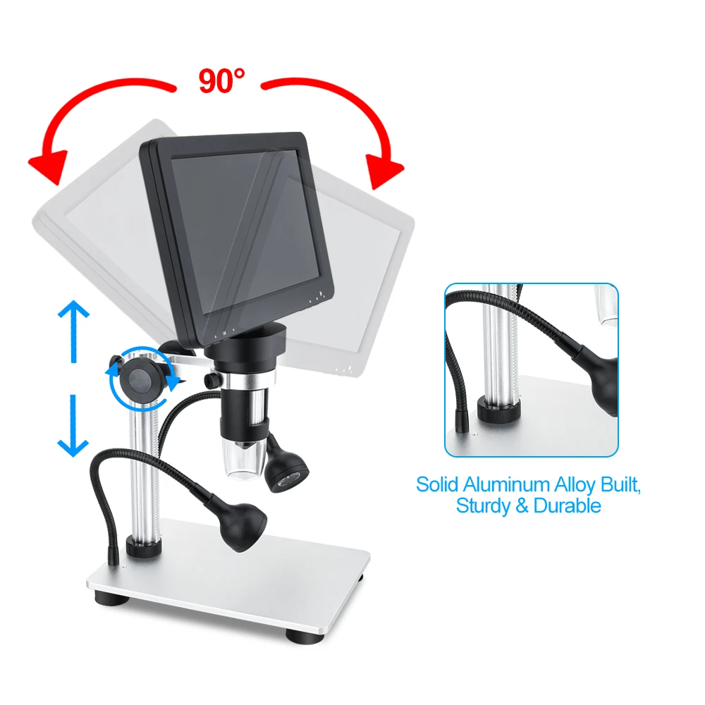
After resting for 5 minutes, place the blood cell counting board on the microscope stage, first use a low-power microscope to locate the location of the counting room, and then switch to a high-power microscope for counting. If the bacterial solution is found to be too thick or too thin before counting, the dilution should be adjusted again before counting. The general sample dilution requirement is that there should be approximately 5-10 bacterial bodies per small cell. Select 5 cells from each counting room (optional 4 corners and center cells) for counting. The mycelium located on the grid line generally only counts on the upper and right lines. If yeast sprouts and the size of the bud reaches half of the mother cell, two bacterial counts are taken. To count a sample, the bacterial content of the sample should be calculated based on the values obtained from two counting chambers.
5. Clean the blood cell counting board
After use, rinse the blood cell counting board with a water column on the water faucet. Do not use hard objects to wash it. After washing, air dry it on your own or blow dry it with a hair dryer. Microscopic examination, observe whether there are residual bacterial bodies or other precipitates in each small cell. If it is not clean, it must be washed repeatedly until it is clean.