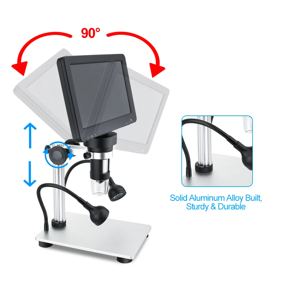
The structure and main components of a fluorescence microscope
(1) Light source
Nowadays, a 200W ultra-high pressure mercury lamp is commonly used as the light source. It is made of quartz glass, with a spherical shape in the middle and a certain amount of mercury filled inside. During operation, the discharge between the two electrodes causes the mercury to evaporate, and the pressure inside the sphere rapidly increases. When the mercury completely evaporates, it can reach 50-70 standard atmospheric pressures, which generally takes about 5-15 minutes. The luminescence of ultra-high pressure mercury lamps is the result of the discharge between electrodes, which continuously dissociates and reduces mercury molecules and emits photons. It emits strong ultraviolet and blue violet light, which is sufficient to excite various fluorescent substances, therefore, it is widely used in fluorescence microscopes.
Ultra high pressure mercury lamps also emit a large amount of heat energy. Therefore, the lamp room must have good heat dissipation conditions and the working environment temperature should not be too high.
The new ultra-high pressure mercury lamp does not require high voltage to ignite in the early stage of use. After some time of use, it needs to be started with high voltage (about 15000V). After starting, the working voltage is generally maintained at 50-60V, and the working current is about 4A. The average lifespan of a 200W ultra-high pressure mercury lamp is about 200h when used for 2 hours each time. The shorter the operating time, the shorter the lifespan. If operated for only 20 minutes once, the lifespan is reduced by 50%. Therefore, try to minimize the number of starts when using. The light efficiency of a light bulb gradually decreases during use. After the light goes out, it needs to wait for cooling before restarting. After lighting the light bulb, it should not be immediately turned off to avoid incomplete evaporation of mercury and damage to the electrode. Generally, it needs to wait for 15 minutes. Due to the high pressure and strong ultraviolet radiation of the ultra-high pressure mercury lamp, the light bulb must be placed in the lamp chamber before being ignited to avoid eye injury and explosion during operation.
The circuit of an ultra-high pressure mercury lamp (100W or 200W) light source and its components including voltage transformation, current suppression, and start-up. There is a system for adjusting the light center of the bulb in the lamp room, with an aluminum plated concave reflector installed behind the bulb bulb and a light collecting lens installed in the front.
The domestically produced ultra-high pressure mercury lamp GCQ-200 has good performance and can replace imported bulbs such as HBO-200, with an average lifespan of over 200 hours and a relatively low price.
A simple and portable high color temperature bromine tungsten fluorescent light source device developed in China, with small volume, light weight, low power, dual use of AC and DC (with built-in DC power supply), easy to carry, convenient to use, has been promoted and applied.
(2) Color filtering system
The color filter system is an important part of a fluorescence microscope, consisting of an excitation filter plate and a compression filter plate. The model of the filter plate is often inconsistent among manufacturers. Filter plates are generally named after the basic color tone, with the first letter representing the color tone, the second letter representing the glass, and the number representing the model characteristics. Olympus microscope
(3) Objective lens
Various objective lenses can be applied, but using achromatic objective lenses is suitable for fluorescence due to their extremely low self fluorescence and transmittance performance (wavelength range). Due to the fact that the fluorescence brightness of the image in the microscope field of view is directly proportional to the square of the objective lens aperture ratio and inversely proportional to its magnification, in order to improve the brightness of the fluorescence image, an objective lens with a larger aperture ratio should be used. Especially at high magnification, its impact is very significant. Therefore, for specimens with insufficient fluorescence, an objective lens with a high aperture rate should be used, combined with an eyepiece as low as possible (4 x, 5 x, 6.3 x, etc.).
(4) Reflective mirror
The reflective layer of a reflector is generally aluminum plated because aluminum absorbs less ultraviolet and visible light in the blue purple region, with a reflection of over 90%, while silver only reflects 70%; Generally, flat reflectors are used.
(5) Spotlight mirror
The concentrator designed and made specifically for fluorescence microscopy is made of quartz glass or other glass that transmits ultraviolet light. There are two types of dark field spotters with distinct field of view. There is also a differential fluorescence concentrator.
(6) Falling light device
The new type of falling light device reflects the shorter wavelength parts (ultraviolet and purple blue) from the light source to the interference spectrophotometer filter due to the properties of the coating on the filter. When the filter is facing the light source at a 45 degree angle. When tilted, it is directed vertically towards the objective lens and directed towards the specimen through the objective lens, causing the specimen to be excited. At this point, the objective lens directly acts as a condenser. At the same time, the long parts of the filter (green, yellow, red, etc.) are transparent to the filter, so they do not reflect in the direction of the objective. The filter acts as an excitation filter plate, and because the fluorescence of the specimen is in the visible light long wavelength region, it can be observed through the filter and reach the objective. The brightness of the fluorescence image increases with magnification, and is stronger than the transmitted light source at high magnification. In addition to its function as a transmissive light source, it is more suitable for direct observation of opaque and semi transparent specimens, such as thick plates, filter membranes, colonies, tissue culture specimens, etc. In recent years, newly developed fluorescence microscopes often use falling light devices, known as falling light fluorescence microscopes.