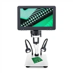
Microscopy - Structure Function Description
objective lens
The objective lens is the optical part of the microscope for the first imaging, which is composed of multiple groups of lenses glued together. The focal length is the total focal length of the lens group.
Depending on the degree of correction for chromatic aberrations, aberrations, field curvature, etc., and their proprietary characteristics, there are several types of objectives: (plan) achromatic objectives, (plan) apochromat objectives, super-plan and specialty objectives, etc.
Chromatic aberration: The color difference in the imaging of visible light sources (polychromatic light). A white object point cannot form a white image point, but a colored image spot.
Aberration: the diffuse spot (circle of confusion) formed at the image plane after the light beam emitted by the object point outside the optical axis is refracted by the optical system.
Coma aberration: Comet-like asymmetric imaging error after the light beam emitted by an object point outside the optical axis is refracted by the optical system.
Achromatic objective lens achromatic objective: ordinary objective lens, marked with "Ach" on the shell. Mainly correct the chromatic aberration (red, blue), spherical aberration (yellow, green) and coma of optical axis imaging. The field curvature is large.
Apochromatic objective lens apochromatic obiective: high-grade objective lens with precise and complex structure, made of special glass such as fluorite, and marked with "Apo" on the shell. On the basis of the achromatic objective lens, it also needs to correct the secondary spectrum, the red, green, and blue aberration, and the red and blue spherical aberration. The apochromatic objective lens has perfect aberration correction, larger numerical aperture, higher resolution, higher effective magnification, and superior imaging quality.
Semi apochromatic objective lens: performance cost and imaging quality are between achromatic objective lens and apochromatic objective lens, also known as fluorspar (fluorite) objective lens, marked with "FL". Chromatic aberration and spherical aberration of red and blue can be corrected.
Plan objective: It mainly corrects the defects of field curvature, so that the field of view is flat, the imaging is realistic, and the observation is convenient. It is a semicircular rear lens added to the objective lens assembly. It can also be combined in achromat objectives, apochromat objectives.
Special objective lens: On the basis of the above objective lens, the objective lens is designed and manufactured to achieve special observation effect.
eyepiece
The eyepiece magnifies the real image of the objective lens, which is the magnification of the intermediate image and belongs to the second magnification. The structure of the eyepiece is relatively simple, consisting of several lenses in several groups. The point where the light rays passing through the eyepiece intersect above is called the eye point, which is the best position for imaging observation.
The eyepiece has a variety of configurations of magnification, 10X is the most commonly used; 5X has higher imaging reducibility, but the magnification is relatively small; 20X eyepiece has the largest magnification, but the image clarity is reduced. It needs to be selected according to actual needs.
Condenser
The condenser is used to make up for the lack of light, appropriately change the light properties of the light source, focus the sample, and improve the illumination. It is located under the stage and must cooperate with it when using NA≥0.40 objective lens. It has a variety of structures, and different numerical apertures of the objective lens have different requirements for the condenser.
1. Abbe condenser (Abbe condenser): Abbe condenser is composed of two lenses, which has better light-gathering ability. When the NA of the objective lens of the ordinary microscope is ≥0.60, the correction of chromatic aberration and spherical aberration is not complete, and it needs to be used together.
2. Achromatic aplanatic condenser: Achromatic condenser is composed of a series of lenses, which can correct chromatic aberration and spherical aberration to obtain satisfactory imaging. It is the best one in bright field observation. It is equipped with advanced microscope and low-magnification objective lens not applicable.
3. Other condensers refer to condensers used for purposes other than the above-mentioned bright field, such as dark field condensers, phase contrast condensers, polarizing condensers, differential interference condensers, etc.
lighting method
Microscope illumination methods are divided into two categories: transmitted illumination and epi-illumination according to the position of the light source and the direction of the beam.
1. Transmitted illumination (transparent illumination) Transmitted illumination is suitable for transparent or translucent samples, and most biological microscopes belong to this type of illumination. Among them, there are two forms of center lighting and oblique lighting.
(1) Central illumination means that the central axis of the illumination beam and the optical axis of the microscope are on the same straight line, which is the most commonly used transmissive illumination method. This method is divided into critical lighting and Kohler lighting.
1) Critical illumination, general illumination method. Advantages: The beam of the light source is imaged by the condenser and irradiated on the sample, and the beam is narrow and strong. Defects: The filament image of the light source coincides with the sample plane, the imaging illumination is uneven, and there is a difference between light and dark. Elimination: Place a milky white heat-absorbing color filter in front of the light source to make the lighting more uniform, or replace the LED light source.
2) Kohler illumination, named in honor of the "secondary imaging" invented by Zeiss engineers. It overcomes the shortcoming of critical illumination, has good imaging effect and good photomicrograph. The main features are: after the filament of the light source passes through the condenser and the variable field of view diaphragm, the filament image falls on the plane of the aperture of the condenser for the first time, and the condenser forms a second filament image at the back focus plane there. The thermal focus is no longer on the plane of the sample, and the sample can be observed with long-term illumination.
(2) oblique illumination (oblique illumination), the central axis of the beam does not coincide with the optical axis of the microscope, and the sample is obliquely illuminated at a certain angle. It is commonly used in phase contrast, dark field, and stereo microscopes.
2. Incident illumination: Incident illumination is also called reflective illumination. The light source is above the sample, and the light beam falls on the sample after passing through the objective lens. The objective lens acts as a condenser and is suitable for non-transparent samples. Fluorescence, stereoscopic, inverted, and confocal microscopes employ this illumination.
Optical axis adjustment
In the microscope optical system, the optical axis of the light source, condenser lens, objective lens, and eyepiece and the center of the diaphragm must coincide with the optical axis of the microscope, and the adjustment of the optical axis before use cannot be ignored.
1. Adjustment of the center of the condenser The adjustment of the center of the condenser is the focus of the adjustment of the microscope optical axis. Method: first reduce the field diaphragm and observe with a 10× objective lens. If the contour image of the diaphragm is not in the center, adjust the two screws on the outside of the condenser to adjust it to the center; then slowly increase the field diaphragm until the contour image is in line with the center. The edges of the field of view coincide, indicating that they are already coaxial, and it is better to use a slightly larger one.
2. Aperture Diaphragm Adjustment The aperture diaphragm is installed in the condenser. There are scale marks on the outer edge of the condenser of the research-grade microscope, which is convenient for adjusting the condenser to match the numerical aperture of the objective lens. It needs to be adjusted synchronously when replacing the objective lens.